Precision Imaging
Excellent cancer treatment and screening outcomes depend upon excellent imaging and biopsy technology. I have spent my career acquiring the best imaging systems available in the world today, including not only systems from Europe, but the best systems made here in the United States. Our imaging systems allow us to visualize and biopsy findings that were invisible on the best systems five years ago. This means that we can detect cancer at an earlier stage as well as clearly identify benign findings without the need for biopsy.
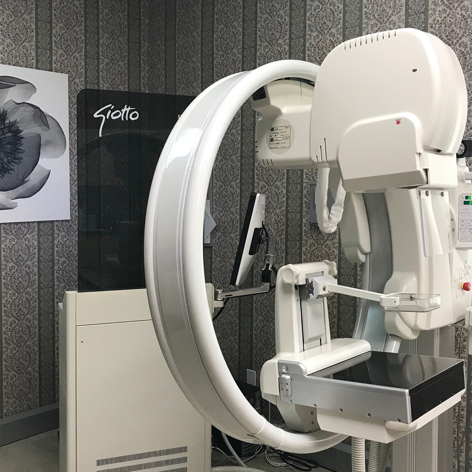
Giotto full-field digital mammography and image-guidance system. Very high-resolution images at half the radiation dose.
Giotto full-field digital mammography
The Giotto full-field digital mammography system allows for excellent image quality at a very high-resolution. One of only four units in the United States, our Giotto produces images of exceptional clarity and quality with half of the radiation exposure of other mammography systems. The system allows for mammograms to be obtained while either standing or sitting. Rotation of the gantry facilitates imaging of more tissue, especially in difficult-to-image areas, improving diagnostic accuracy.
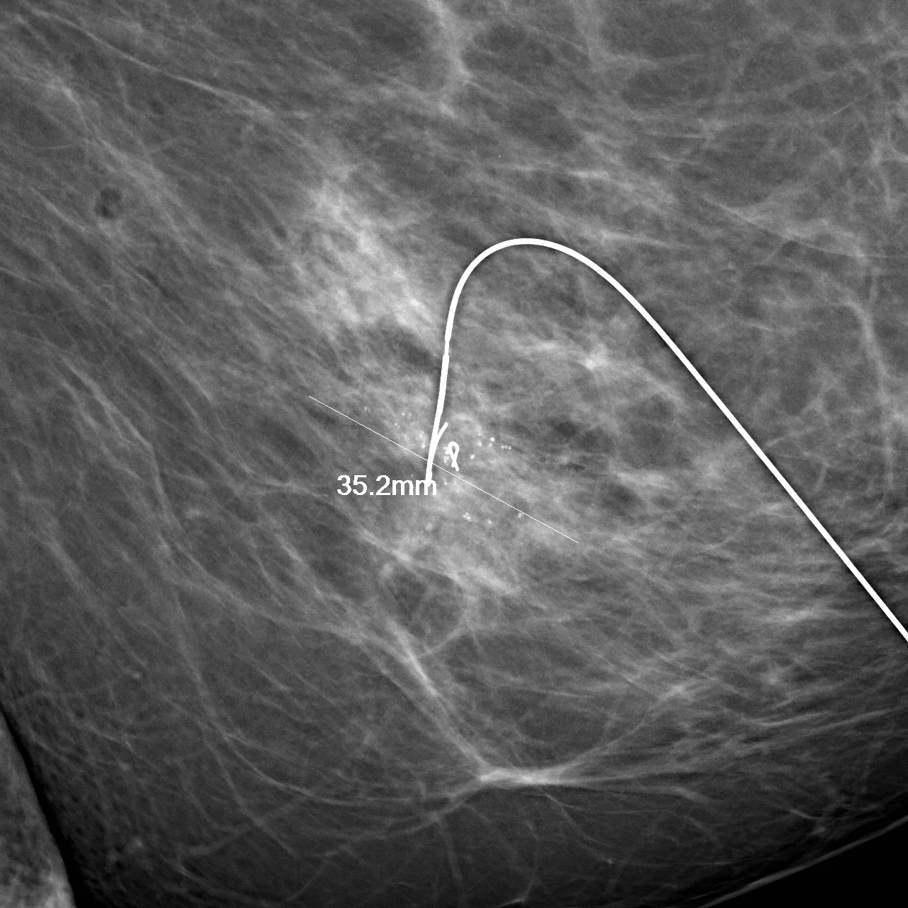
Surgical localization and planning on the Giotto. Direct measurement of the cancer and placement of localization devices with millimetric precision decreases reexcision rates while minimizing the amount of tissue that has to be removed.
Giotto digital multiplanar imaging guidance
Designed not just for imaging, but for imaging guidance, our Giotto mammography system allows for the easy localization and biopsy of any finding that we can see on mammogram. This includes small areas of microcalcifications as well as mass lesions. Biopsies can be performed in the position of the client's best comfort.

This client has an 11mm breast cancer with a 7mm satellite cancer 27mm away. Mapping with the Invenia allowed both lesions to be removed with one lumpectomy incision. The smaller lesion was not seen on mammogram.
Invenia whole-breast sonographic tomography
The Invenia whole-breast ultrasound system images the entire breast and axilla in comfort. Spectacular resolution allows the detection of small cancers missed by mammography. The system is also useful for diagnostic evaluation of breast lumps and mapping the extent of disease prior to surgery. About 25% of the cancers we detect are not seen on mammogram, but are detected only on the Invenia.
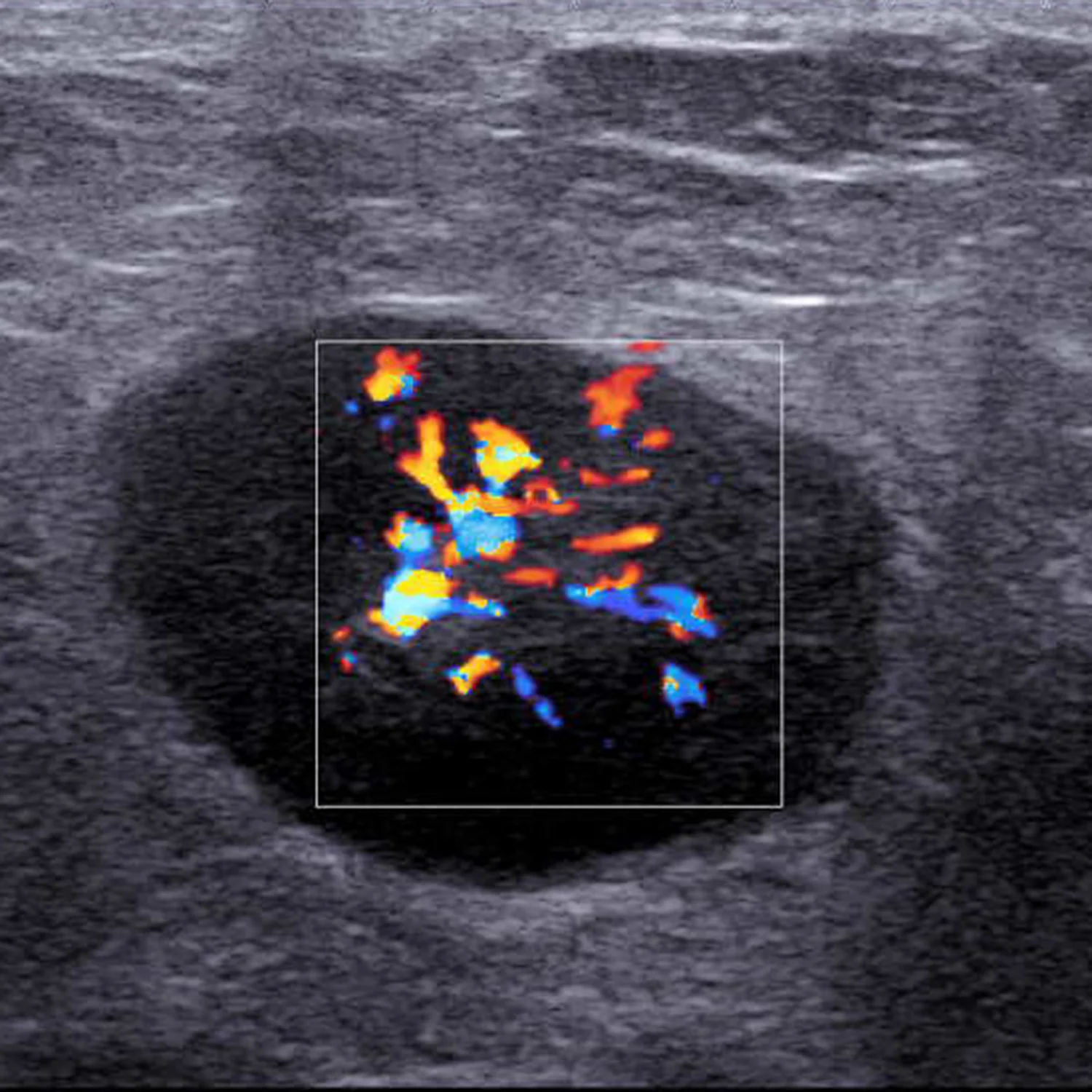
Color Doppler imaging of a malignant lymph node in a 55-year-old client with breast cancer.
AIX Explorer high-resolution focus ultrasound
For detailed imaging of an abnormal finding, as well as biopsy guidance, we use the AIX Explorer ultrasound system. The AIX system employs color and power Doppler flow imaging to assess blood flow as well as shear-wave supersonic tissue imaging to determine tissue stiffness. These technologies better characterize a finding, allowing the safe avoidance of biopsy for benign findings.
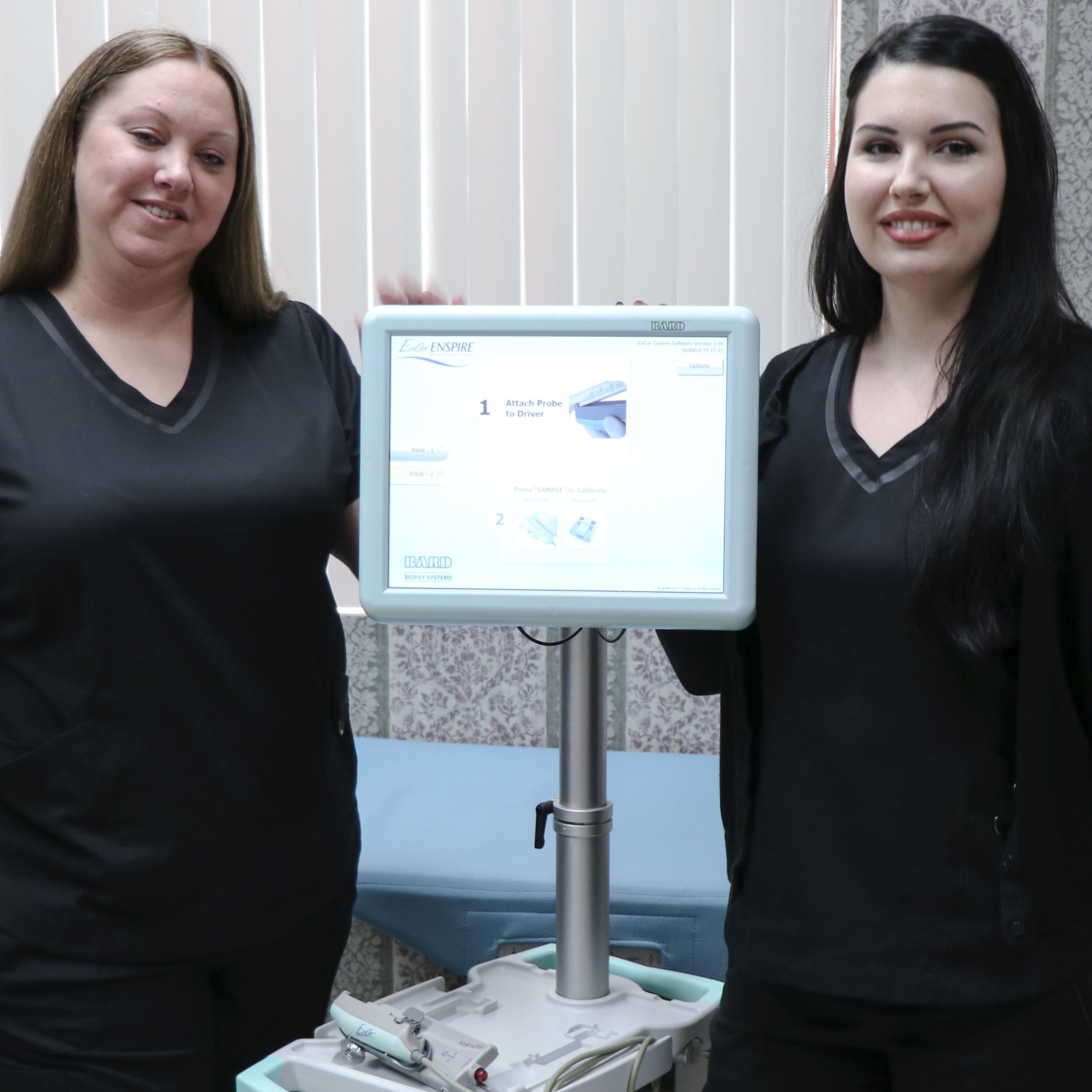
Bard Encor vacuum-assisted biopsy instrument
The Bard breast biopsy instrument is used in stereotactic, or mammogram-guided biopsy to remove small lesions, especially areas of calcifications. Often small areas of calcifications can be removed using the Bard system, minimizing the need for further follow-up imaging.